

Atlas of Medical Mycology – Reena Ray Ghosh
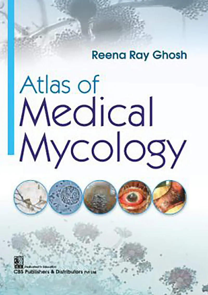
Atlas of Medical Mycology by Reena Ray Ghosh is a practical visual resource designed to bridge the gap between detailed medical mycology textbooks and the day-to-day requirements of the laboratory. Rather than relying exclusively on lengthy theoretical explanations, the atlas uses photographs and concise descriptions to help students and laboratory learners recognize and appreciate the characteristic features of medically important fungi.
The book showcases culture morphology photographs and photomicrographs of both common and uncommon fungi isolated in the Mycology Laboratory Division of RG Kar Medical College, Kolkata. The accompanying descriptions are designed to help readers develop the visual recognition skills needed for laboratory identification, while clinical photographs and brief case descriptions provide an additional connection between laboratory findings and patient presentation.
About the Book
A Visual Introduction to Medical Mycology
Recognizing fungi in the laboratory requires familiarity with their appearance in culture and under the microscope. Atlas of Medical Mycology provides a visual approach that complements conventional medical mycology textbooks and helps learners become more comfortable with fungal morphology.
The photographs provide an opportunity to observe relevant features directly rather than relying solely on written descriptions.
Culture Morphology
The atlas includes photographs showing the culture morphology of fungi, providing valuable visual reference material for students learning how fungal isolates can appear in laboratory culture.
The inclusion of both common and uncommon fungi broadens the reader’s exposure to the range of organisms that may be encountered in medical mycology.
Photomicrographs and Microscopic Features
Microscopic examination is an important part of fungal identification. The book includes photomicrographs accompanied by pertinent descriptions that draw attention to relevant morphological characteristics.
This combination of images and explanatory text is particularly useful for learners who are still developing the ability to distinguish important fungal features under the microscope.
Clinical Photographs and Case Descriptions
The atlas extends beyond laboratory morphology by including clinical photographs and brief case descriptions associated with the fungal isolates. These cases help students understand how laboratory findings relate to actual clinical presentations.
By connecting the patient, clinical findings, and fungal isolate, the book encourages the development of clinical reasoning alongside laboratory skills.
Common and Uncommon Fungi
Medical mycology students need exposure to both frequently encountered organisms and less familiar fungal isolates. The atlas presents photographs of common as well as uncommon fungi, providing a broader visual reference for laboratory learning.
Bridging Textbooks and the Laboratory
One of the principal purposes of the book is to serve as a bridge between the extensive theoretical information found in medical mycology textbooks and the practical experience of examining fungal isolates.
Its concise descriptions and extensive visual material make it a useful supplementary resource for students beginning practical work in medical mycology.
Who Should Use This Book?
- Medical students studying medical mycology
- Students learning clinical microbiology
- Medical laboratory technology students
- Clinical microbiology laboratory trainees
- Students developing fungal identification skills
- Laboratory professionals working with fungal isolates
- Teachers and instructors of medical mycology
- Readers seeking a visual reference for medically important fungi
Final Thoughts
Atlas of Medical Mycology offers a practical and visually oriented way to study medically important fungi. Its combination of culture photographs, photomicrographs, clinical images, and concise descriptions helps learners develop the visual skills needed to recognize fungal morphology while also appreciating its clinical significance.
For students moving from theoretical medical mycology into laboratory practice, the atlas provides a useful companion to conventional textbooks. The inclusion of clinical cases further strengthens the connection between fungal identification, laboratory findings, and patient presentation.
Book Information
Title: Atlas of Medical Mycology
Author: Reena Ray Ghosh
Publisher: CBS Publishers and Distributors Pvt Ltd
Publication Date: January 30, 2019
Edition: First Edition
Language: English
Print Length: 200 pages
ISBN-13: 978-8194116783
ASIN: B07SPBR1ZL
Subjects: Medical Mycology, Clinical Mycology, Fungal Identification, Fungal Morphology, Culture Morphology, Photomicrography, Diagnostic Mycology, Clinical Microbiology, Laboratory Diagnosis, Medical Laboratory Science
Atlas of Medical Mycology is a practical visual guide that connects fungal culture morphology and microscopy with clinical photographs and case-based information to support the study and laboratory identification of medically important fungi.
Support the Creators!
If you enjoy this book, please consider supporting the author and publisher by purchasing a physical or digital copy. Buying official copies ensures authors can keep writing great books.