
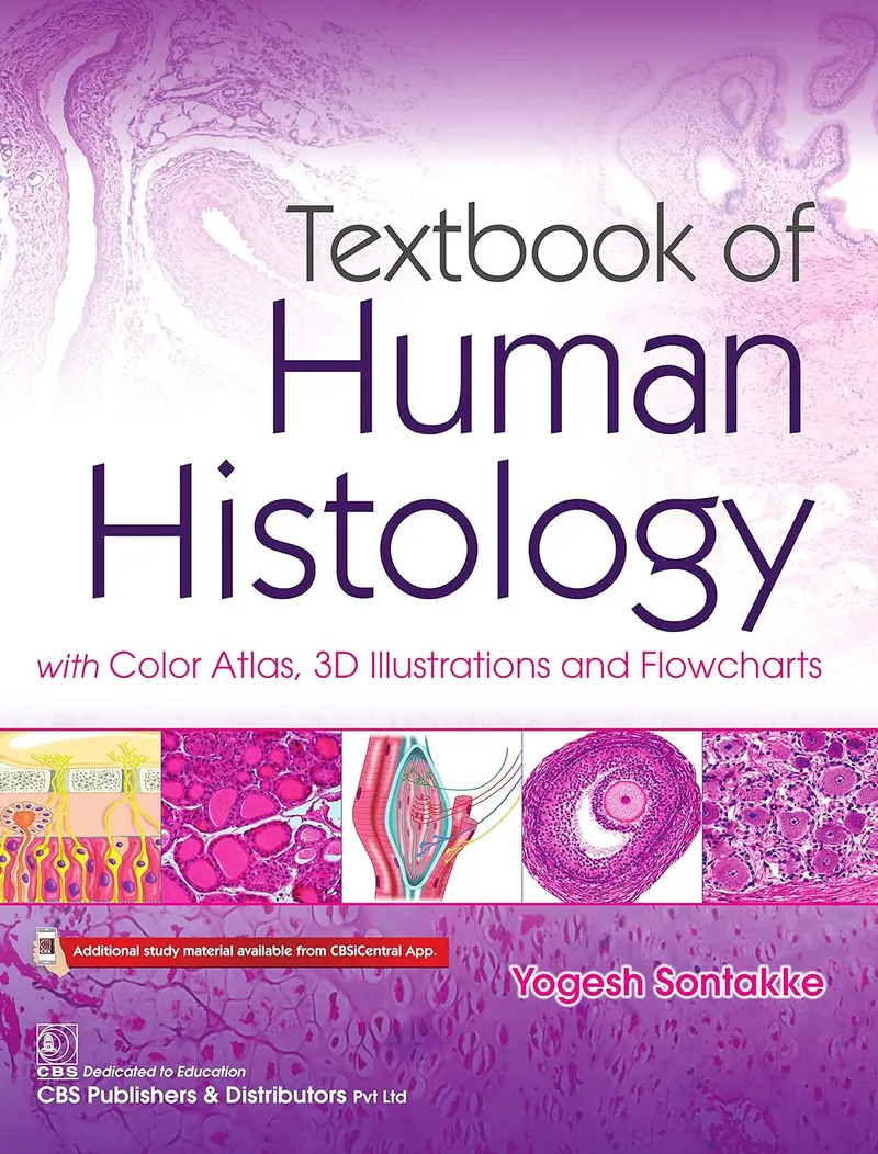

Textbook of Human Histology with Color Atlas, 3D Illustrations and Flowcharts
Textbook of Human Histology with Color Atlas, 3D Illustrations and Flowcharts is a comprehensive and student-friendly resource designed primarily for undergraduate medical students. The book presents the essential concepts of human histology in a concise format while combining traditional histology content with extensive visual learning tools.
With photomicrographs, flowcharts, practice figures, and 3D illustrations, the book helps students understand and recognize microscopic structures while making complex concepts easier to revise and remember. Functional and clinical correlations further connect microscopic anatomy with the structure and function of the human body and the development of disease.
About the Book
The book is designed around the practical needs of medical students, particularly those preparing for examinations. Its concise explanations allow students to quickly review important histological concepts, while the visual resources provide additional support for understanding and memorizing microanatomy.
The inclusion of an examination-focused summary, reproducible practice figures, and clearly separated interesting facts makes the book useful both for learning new material and for rapid revision before assessments.
Key Features
Color Atlas and Photomicrographs
The book includes 117 photomicrographs that help students recognize microscopic structures and develop the visual skills required for practical histology examinations.
Flowcharts for Quick Revision
With 122 flowcharts, important concepts and relationships are presented in a simplified visual format. These flowcharts can make large amounts of information easier to organize, recall, and revise.
Practice Figures
The book provides 106 practice figures in H&E pencil-drawing style. These illustrations are designed to be easily reproduced in theory examinations, helping students practice presenting histological structures clearly.
3D Illustrations
A collection of 175 three-dimensional illustrations provides a visual approach to difficult concepts. These illustrations can help bridge the gap between two-dimensional microscopic images and the three-dimensional organization of tissues and structures.
Examination Guide
A dedicated summary section provides an examination-oriented review of important facts. This allows students to consolidate key information and prepare more efficiently for written assessments.
Interesting Facts
Selected interesting facts are separated from the main text so that additional information can be easily identified without interrupting the flow of the core material.
Clinical Correlations
Clinical correlations provide a connection between normal histological structure and disease processes. This vertical integration helps students understand the relevance of histology to pathology and clinical medicine.
Who Should Use This Book?
- Undergraduate medical students studying human histology
- Students preparing for histology theory and practical examinations
- Medical students who prefer visual learning resources
- Learners reviewing microscopic anatomy and tissue organization
- Students studying the relationship between histology and pathology
- Teachers and instructors involved in undergraduate medical education
Final Thoughts
Textbook of Human Histology with Color Atlas, 3D Illustrations and Flowcharts combines concise explanations with a large collection of visual and examination-oriented resources. Its photomicrographs, flowcharts, practice drawings, and 3D illustrations provide multiple ways for students to understand and retain difficult histological concepts.
By incorporating functional and clinical correlations, the book also demonstrates how microscopic anatomy contributes to understanding disease and clinical practice. It is therefore a useful study and revision companion for undergraduate medical students throughout their histology coursework.
Book Information
Title: Textbook of Human Histology with Color Atlas, 3D Illustrations and Flowcharts
Publisher: CBS Publishers and Distributors Pvt Ltd
Publication Date: August 29, 2020
Language: English
Print Length: 356 pages
ISBN-13: 978-9354661358
ASIN: B08GZ3H6YF
Subjects: Human Histology, Histology, Microscopic Anatomy, Medical Histology, Tissue Biology, Color Atlas, Medical Education, Histology Practical, Histology Examination, Pathology
Textbook of Human Histology with Color Atlas, 3D Illustrations and Flowcharts is a visually rich histology resource combining photomicrographs, flowcharts, practice figures, 3D illustrations, and clinical correlations for undergraduate medical students.
Support the Creators!
If you enjoy this book, please consider supporting the author and publisher by purchasing a physical or digital copy. Buying official copies ensures authors can keep writing great books.